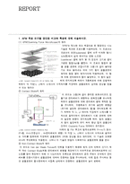
Mode의 원리
이 모드는 그림2와 같이 분리된 포토다이오드 검출기로 캔티레버가 편향되는 변화정도를 모니터링하면서 샘플표면에 대해 캔티레버 끝에 부착된 팁을 주사하여, 귀환회로가 초기에 설정한 편향값 (초기에 설정한 캔티레버의 휨)을 유지하도록 하기 위하여 각 (x, y) 데이터 지점에서 스캐
scanning a focused electron beam across the surface of a specimen. In the most common mode, the low energy secondary electrons emitted are detected and used to modulate the brightness of a synchronously scanned CRT. Other signals can also be detected. X-rays, characteristic of that part of the specimen probed by the electron beam, allow both a qualitative and quantitative determination of the ele
recognition in nanoscale systems.
While microfabrication techniques such as photolithography, microcontact
printing, micromachining, and microwriting can produce patterns as small
as 0.1 mm, production of sub-100-nm structures still poses a significant
challenge.
At present, such high-resolution fabrication can be achieved using scanning
probe lithography (SPL).
투과전자현미경은 주로 시료의 내부구조나 단면을 관찰하는데 쓰이고 있다. 원리는 광학현미경과 비슷하다. 전자현미경에서의 광원은 높은 진공 상태(1x10⁻⁴ 이상)에서 고속으로 가속되는 전자선으로 이 전자선이 표본을 투과하여 형광판이나 사진필름에 초점을 맞추어 투사된다. 이 전자의 파장은 가