infiltrating inflammatory cell
Luminal narowing, fibrosis로 기도저항 증가
Parenchyma
Alveolar septum이 파괴, airspace 확장
Bronchoalveolar lavage fluid from smoker
Macrophage 증가(>95% of total cell count)
Neutrophil 1~2%(비흡연자에서는 거의 없음)
Clinical presentation
History
3대 증상 : 기침, 가래, 호흡곤란
기침
COPD 환자의 가장 흔한 증상
대부분
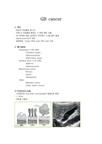
infiltrating type, 담낭벽의 미만성 비후를 보이는 침윤 형
IV massive tumor type, 담낭 전체가 종양으로 점거된, 결석을 동반한 type
V large solitary type, 담낭 전체가 큰 단일의 종양으로 형성된 type
4. 원인
발생기전은 아직 unknown, 유전적/환경적 요인 추정
5. 위험인자
a. GB stone의 risk factor와 비슷 : 90%에서
infiltrate comprised primarily of T-lymphocytes [76,77]. This infiltrate is not diagnostic, and it may be seen in a wide variety of conditions, including a simple drug-induced exanthem. A sparse infiltrate of lymphocytes develops at the dermoepidermal junction, with lymphocytes clustered around dying basal keratinocytes ("satellitosis") [9]. As the lesions progress, frank subepidermal vesiculatio
Repeat biopsy at 6 monthes
: Strong predictor of ESRD
- ongoing inflamations with cellular crescent, macrophages in the tubular lumens, immune deposits
Membraneous > Proliferative
Pure membranous > Superimposed Proliferative lesions( Class III + V , Class IV +V)
- 72% 10-year survival rates vs 20-48% 10-year survival rates
aggressive treatment to avoid irrevers