Ⅰ. 서론
우리는 4월 27일 외래를 통해 입원한 김00(F/69) 환자를 만나게 되었다. 간호정보 사정을 위해 김00님의 병실을 방문한 후 입원 동기를 물어보니 혈관이 부풀어 오르고 크기가 더 커져서 수술을 하기 위해 내원하셨다고 한다. 주요 간호기록지 정보로는 교육수준이 초졸 이하이며 입원동기로는
aorticdissection 받은 환자에게 행하는 장기적인 치료:
beta blockers 나 다른 antihypertensive agents(ACE inhibitors 나 calcium antagonist)를 사용하여 hypertension 조절 과 cardiac contractility 억제
detect propagation or expansion을 감지하기 위해 외래에서 6-12달 마다 contrast-enhanced CT or MRI가 시행되어야 한다.
-dissections을
Widening of the mediastinum on an x-ray of the chest has moderate sensitivity (67%)
The calcium sign
Pleural effusions
Magnetic resonance imaging (MRI) is currently the gold standard test for the detection
right atrial appendage
2) 원인별 병리해부소견
(1) 동맥경화성 동맥류(arteriosclerotic aneurysm)
① 흉부 대동맥류의 가장 흔한 원인
② 하행 흉부대동맥에서 가장 빈발 > 상행 흉부대동맥 > 대동맥궁(가장 적음)
③ 모양은 보통 방추상(fusiform)이나 주머니 모양
(2) 대동맥박리(aorticdissection)
① 대동맥류의 두 번째 많은 원인
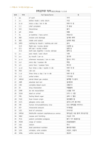
1 aa of each 각각
2 a.c before meals / ante cibum 식전
3 b.i.d twice a day / bis in die 하루 두 번
4 C.C chief complaint 주 증상
5 D/C Discontinue 중지
6 gtt drops 방울
7 hs at bedtime / hora somni 취침시간
8 I&D incision and drainage 절개와 배액
9 I&O intake and output 섭취와 배설
10 NPO nothing by mouth / nothing per oral 금식
11Right eye / oculus dexter