증상이 악화되어 추적 검사 상 혈종이 확인되었을 때를 말한다.
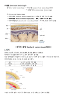
2. 해부학적 구조
<Brain>
- 무게: 1300~1600g
- 기능: 모든 생각, 보는 것, 말하는 것, 냄새맡는 것, 감정, 또한 우리 몸이 움직이기
위해 필요한 모든 행동을 관장함
- 이처럼 중요한 기능을 하는 뇌는 여러 막으로 보호되어 있음.
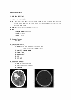
midbrain과 right basal ganglion의 lenticular nucleus에 hemorrhage가 관찰됨
-> a moderate amount of IVH & ICH at the midbrain & medial aspect of right
lentiform nucleus
- brain angio CT & 3D : unremarkable CTA of brain and unremarkable enhanced
brain
->Hypertensive intracerebral hemorrheage
4. 향후 Plan & 보호자에게 말해 줄 수 있는 Complication
2. ER에서 해야 할 처치 :
1) ABC유지 : air way, breathing, circulation 유지
ABGA f/u 하면서 인공호흡기 삽입
2) Brain CT, C-spine lateral
3) Chest X ray
4) Abdominal CT
5) EKG
6) IICP 방지
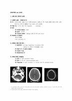
3. Initial Brain Imaging
1) SDH at both frontallobe
2) ICH at right frontal, parietal
3) Fractures
4. 향후 Plan & 보호자에게
1. A crescentic SDH at the Rt. frontoparietal area and hemorrhagic cerebral contusion at the Lt. frontallobe with mild midline shift to the Rt. side.
2. Traumtic SAH at the basal cisterns with diffusion brian swelling.
3. Hemorrhiac cerebral contusion at the Rt. temporoparietal area with a curvilinar SDH at the Rt. temporoparietal area and hemorrhagic cerebral contusion at the Lt. high parieta
1. 다음 중 시야(visual field) 장애 검사법은?
1) corneal reflex
2) confrontation test
3) Doll's eye movement
4) Snellen eye chart test
5) Weber test
답) 2번
1) 5번 신경을 이상을 알아보기 위한거죠.. 반사 회로는 5번으로 들어갔다가 7번으로 나옵니다.
2) 환자가 검사자의 코를 보게 한 후 검사자의 손을 밖에서부터 안으