2. ER에서 해야 할 처치 :
1) ABC : air way 유지, breathing, circulation
2) EKG
3) Brain CT
4) 뇌압감소
-두부의 위치를 높여준다(30~45‘)
-과호흡 (pCO2 30~35기준)
-mannitol
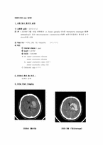
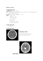
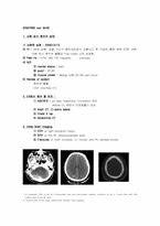
3. Initial Brain Imaging
1) A large ICH at the right basal ganglia and periventricualar white matter with large amount of IVH.
4. 향후 Plan & 보호자에게 말해
4. 향후 Plan & 보호자에게 말해 줄 수 있는 Complication
1) Plan : PHL with microscopic L-discectomy Lt. L4-5
2) Complication :
- 일반적인 수술의 합병증 : 출혈, 감염, 신경의 손상, thrombophlebitis,
지속적 동통
5. 1st OP
1) OP date : 2010/2/24
2) OP name : microscopic L-discectomy Lt. L4-5
3) OP 한 이유 : 지속적인 동통, 운동
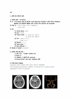
진단: Acute Traumatic SDH
4. 향후 Plan & 보호자에게 말해 줄 수 있는 Complication
Plan: Emergent Operation
ICP가 증가되어 Cerebral Hernia에 의한 증상: Anterior Cerebral artery와 Corpus callosum 손상
수술에 의한 합병증: 감염- 수막염 및 뇌 농양
5. 1st OP
OP date : 2010/02/27
OP name : craniectomy,Lt
2월2일 Corpus callosum의 splenium 근처에 약 2.7 x 1.8 x 2.7cm size의 tangled vascular structure가 관찰되며 feeding artery는 ACA(pericallosal a.)와 right PCA로 보이면서 draining vein은 enlarged되어 있고 vein of Galen이나 straight sinus로 drainage됨.
Angio : AP view상에서는 3 x 2cm크기의 AVM. A small AVM at the splenium of corpus callosum and pericallosal area
4. 향후 Plan & 보호자에게 말해 줄 수 있는 Complication
- IV urokinase for thrombolysis
- Complication
뇌출혈
기타 장기의 출혈
출혈로 인한 사망
조영제에 의한 s/e
색전증에 의한 다른 문제
5. IA uk 후 problem
- 환자 시술 당일 저녁 mental change 보여서 f/u CT 촬영
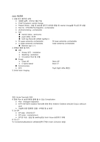
6. F/U brain imaging
<1/19 f/u CT>
2. ER에서 해야 할 처치 :
1) ABC 유지 : 기도확보(suction, intubation), BP정상 유지
2) Brain CT
3) seizure시 anticonvulsant
4) bed rest
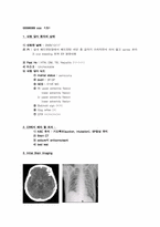
3. Initial Brain Imaging
Conclusion: Subarachnoid hemorrhage due to Rt. posterior communicating artery aneurysmal rupture
4. 향후 Plan & 보호자에게 말해 줄 수 있는 C
3. Initial Brain Imaging
1) angio
2) CT
4. 향후 Plan & 보호자에게 말해 줄 수 있는 Complication
- 뇌혈관조영술, 절대안정, 코일 색전술, 결찰
- complication : 재출혈, 뇌연축, 수두증
- 수술에 대한 complication : vomitting, 경련, shock
5. 1st OP
1) OP date : 2010/02/24 수술예정
2) OP name :
1. A crescentic SDH at the Rt. frontoparietal area and hemorrhagic cerebral contusion at the Lt. frontal lobe with mild midline shift to the Rt. side.
2. Traumtic SAH at the basal cisterns with diffusion brian swelling.
3. Hemorrhiac cerebral contusion at the Rt. temporoparietal area with a curvilinar SDH at the Rt. temporoparietal area and hemorrhagic cerebral contusion at the Lt. high parieta