CT, C-spine lateral
3. Initial Brain Imaging
1) C- spine lat. : fracture in C5 vertebral body with complete dislocation.
2) C- spine CT, 3D
4. 향후 Plan & 보호자에게 말해 줄 수 있는 Complication
1) Plan : Decompressive craniectomy, hematoma 제거
2) Complication
- 척수손상 -> 호흡정지, 심정지
- 척추 신경 손상
3) 수술의 합병증
2. ER에서 해야 할 처치
- ABC 유지
Airway 유지 - intubation
Breathing and ventilation
Circulation
- IICP 방지
head elevation
sedation
osmotic agents
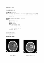
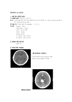
- Brain CT
3. Initial brain imaging
- brain CT : ventricle에 moderate amount의 high density hemorrhage가 관찰되며
midbrain과 right basal ganglion의 lenticular nucleus에 h
4. 향후 Plan & 보호자에게 말해 줄 수 있는 Complication
1) Plan : PHL with microscopic L-discectomy Lt. L4-5
2) Complication :
- 일반적인 수술의 합병증 : 출혈, 감염, 신경의 손상, thrombophlebitis,
지속적 동통
5. 1st OP
1) OP date : 2010/2/24
2) OP name : microscopic L-discectomy Lt. L4-5
3) OP 한 이유 : 지속적인 동통, 운동
2월2일 Corpus callosum의 splenium 근처에 약 2.7 x 1.8 x 2.7cm size의 tangled vascular structure가 관찰되며 feeding artery는 ACA(pericallosal a.)와 right PCA로 보이면서 draining vein은 enlarged되어 있고 vein of Galen이나 straight sinus로 drainage됨.
Angio : AP view상에서는 3 x 2cm크기의 AVM. A small AVM at the splenium of corpus callosum and pericallosal area