OP
1) OP date : 2010/1/18
2) OP name : Ventriculo-peritoneal shunt
3) OP 한 이유 : Enlarged Rt. lateral ventricle
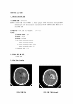
6. F/U CT
1월 19일 1월 26일
7. 2nd OP
1) OP date : 2010/2/1
2) OP name : cranioplasty with bone cement
3) OP 한 이유 : skull defect(Lt.)
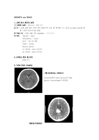
8. F/U CT
2월 2일
Plan: Emergent Operation
ICP가 증가되어 Cerebral Hernia에 의한 증상: Anterior Cerebral artery와 Corpus callosum 손상
수술에 의한 합병증: 감염- 수막염 및 뇌 농양
5. 1st OP
OP date : 2010/02/27
OP name : craniectomy,Lt.
OP 한 이유 : 감압 및 swelling되는 brain tissue 보호하기 위해
6. F/U CT
Tx: Cra
CT Chest & HR Double Study(04.18)
- No evidence of intrathoracic metastasis.
- Comparing to prior chest CT (2010-01-15) : No evidence of metastasis or active parenchymal consolidation in both lung parenchyma. No LNE in the hilar, mediastinum and both supraclavicular fossa. Left thyroid nodule.
8. Impression & Plan
Impression
- rectal cancer
- liv
Plan & 보호자에게 말해 줄 수 있는 Complication
Plan : 색전술후 AVM의 수술적 제거
*색전술로 유입동맥을 초정밀 선택(superselection)하여 뇌동맥기형으로만 가는 혈
류를 일부를 막아 핵의 크기를 줄임으로써, 수술적 제거를 용이하게 한후 수술로 제거
AVM이 뇌실내에 위치해 있고 약 6-18%에서 재출